Ravosa Lab

Research Specialities
Evolutionary Morphology
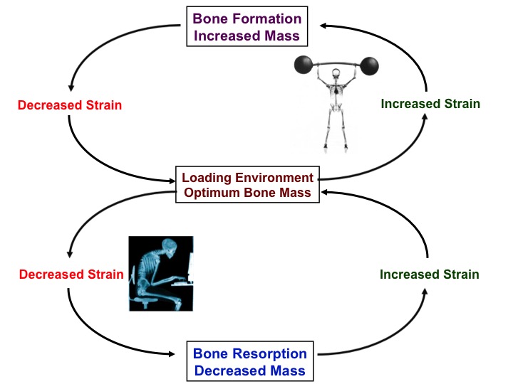
Mechanobiology
Pathobiology
Aging & Development
Musculoskeletal Systems
Skull & Feeding Apparatus
Plasticity
Experimental biology
Evolutionary morphology and pathobiology of the vertebrate skull, masticatory complex and musculoskeletal system
Much like many members of the general public and scientific communities, I grew up fascinated by the astounding phenotypic variability of living and fossil organisms. Mammals are one such group that fails to disappoint. In this clade, the greater diversification and central role of the masticatory apparatus in food procurement and processing has figured prominently in studies of craniomandibular biomechanics and evolution. Indeed, being among the most highly mineralized, and thus well preserved, tissues in the body, craniodental (and postcranial) remains have long been used by paleontologists to offer novel insights into the behavior and phylogenetic affinities of extinct taxa.
Over the past three decades, my integrative research program has investigated major adaptive and structural transformations in mammalian musculoskeletal form during ontogeny and across higher-level clades. With an eye to both the evolutionary and translational implications, our lab has investigated the plasticity, mechanobiology, ecomorphology, aging and performance of the mammalian musculoskeletal system. Work on development, biomechanics and evolution has marshaled diverse and non-traditional sources of evidence to address outstanding issues concerning the complex underpinnings of patterns of phenotypic variation. To this end, we have employed of a broad range of modern cell biological, molecular, engineering and imaging techniques (immunostaining, histomorphometry, tissue properties testing, microarray, tissue culture, microCT, PCR) as well as unique experimental and transgenic animal models.
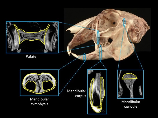
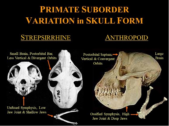
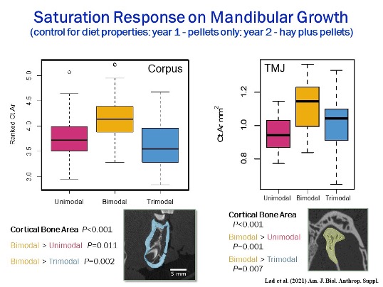
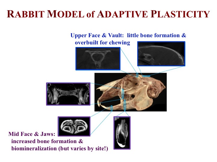
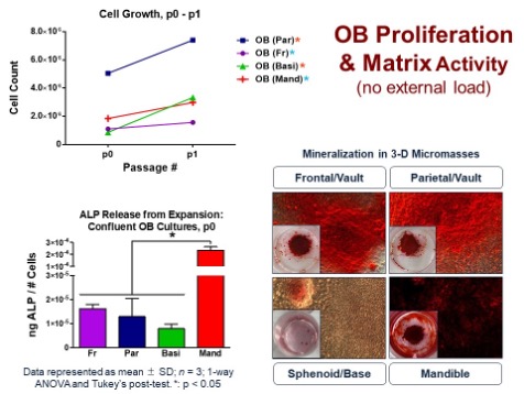
We have developed a rabbit model of dietary plasticity in cranial hard- and soft-tissues, which is being coupled with data on load-induced changes in gene expression patterns of jaw-joint cartilage. This evidence is being integrated with in vivo data regarding the relationship between food mechanical properties and chewing behavior. Such research also contributes to ongoing debate regarding the ecomorphological significance of dietary seasonal variability and daily patterns of feeding modality in cranial evolution among fossil and living organisms. Histomorphometric analyses have been applied to the developing proximal humerus and femur in growing mini-pigs. These first-ever long-term studies have applied a more naturalistic and integrative perspective to the plasticity and performance of multiple cranial and limb joints. Our lab has also investigated the first mouse model of neural encephalization, which has important implications for understanding prenatal development, cranial dysmorphologies and the evolution of the remarkably large brain in humans. These findings are being coupled with in vitro analyses of site-specific variation in the mechanobiology of bone cells, dura mater cells and cartilage cells. Such information will be critical for developing a model of extrinsic and intrinsic determinants of phenotypic diversity in the vertebrate skull. Ongoing macroscale tests of the properties and biomechanics of an ossified mandibular symphysis, a jaw-joint feature characteristic of modern anthropoids, has capitalized on various analytical and technical methods common to engineering. Investigation of the dynamic links among skeletal safety factors, masticatory stresses and cortical bone formation has highlighted critical similarities and differences in loading patterns between vertebrate skulls and limbs. From a clinical and bioengineering standpoint, such information is important for characterizing strain-mediated responses necessary for mimicking the natural growth activity of connective tissues. Finally, we are developing a rabbit model of osteonecrosis of the jaw, a debilitating oral disease associated with long-term bisphosphonate therapy used in treating bone metastases and osteoporosis.
In synthesizing seemingly disparate sources of biological evidence and novel techniques, we have sought to uniquely address a number of difficult and longstanding questions regarding the function, development, pathobiology and evolution of the musculoskeletal system, skull and feeding complex in mammals and other vertebrates.
Education
- Postdoctoral, Duke University Medical Center
- PhD, Biological Anthropology and Anatomy, Northwestern University, 1989
- MA, Biological Anthropology and Anatomy, Northwester University, 1986
- BA, Interdepartmental Studies and Paleoanthropology, University of Rochester, 1983
Research Projects
Selected Publications
Consolini, J., Oberman, A.G., Sayuta, J., Damen, F.W., Goergen, C.J., Ravosa, M.J. & Holland, M.A. (2024) Investigation of direction- and age-dependent prestretch in mouse cranial dura mater. Biomechanics Modeling Mechanobiology 23:doi.org/10.1007/s10237-023-01802-6.
Lad, S.E., Kowalkowski, H., Liggio, D.F., Ding, H. & Ravosa, M.J. (2023) Prolonged cyclical loading induces Haversian remodeling in mandibles of growing rabbits. J. Experimental Biology 226: jeb245942.
Harper, E.I., Siroky, M.D., Hilliard, T.S., Dominique, G.M., Hammond, C., Liu, Y., Yang, J., Hubble, V.B., Walsh, D.J., Melander, R.J., Melander, C., Ravosa, M.J. & Stack, M.S. (2023) Advanced glycation end products as a potential target for restructuring the ovarian cancer microenvironment: A pilot study. International J. Molecular Sciences 24:9804.
Mitchell, D.R., Wroe, S., Ravosa, M.J.& Menegaz, R.A. (2021) More challenging diets sustain feeding performance: Applications towards captive rearing of wildlife. Integrative Organismal Biology 3:obab030. (cover image)
Kraatz, D., Belabbas, R., Fostowicz-Frelik, Ł., Ge, D.-Y., Kuznetsov, A.N., Lang, M., Lopez-Torres, S., Racicot, R.A., Ravosa, M.J., Sharp, A.C., Sherratt, E., Silcox, M.T., Słowiak, J., Winker, A.J. & Ruf, I. (2021) Lagomorpha as a model organism. Frontiers in Ecology and Evolution: Phylogenetics, Phylogenomics, and Systematics 9:ar636402.
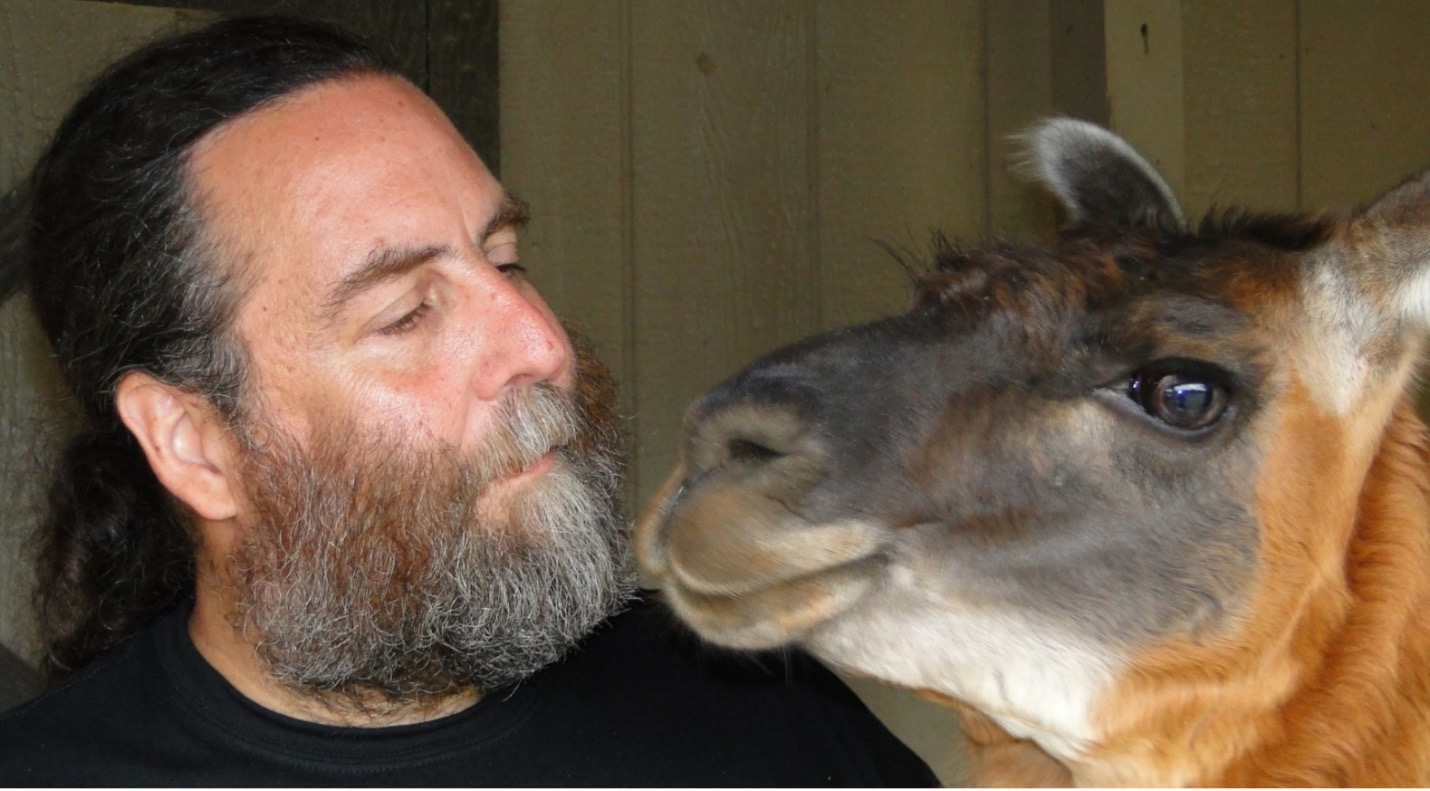
Nett, E.M., Jaglowski, B., Ravosa, L.J., Ravosa, D.D. & Ravosa, M.J.(2021) Mechanical properties of food and masticatory behavior in llamas, Llama glama. Journal of Mammalogy 102:1375-1389.
Lad, S.E., Anderson, R.J., Cortese, S.A., Alvarez, C.E., Danison, A.D., Morris, H.M. & Ravosa, M.J.(2021) Bone remodeling and cyclical loading in maxillae of New Zealand white rabbits (Oryctolagus cuniculus). Anatomical Record 304A:1927-1936.
Terhune, C.E., Sylvester, A.D., Scott, J.E. & Ravosa, M.J.(2020) Trabecular architecture of the mandibular condyle of rabbits is related to dietary resistance during growth. Journal of Experimental Biology 223:jeb220988. (cover image)
Ravosa, M.J.& Vinyard, C.J. (2020) Masticatory loading and ossification of the mandibular symphysis during anthropoid origins. Nature Scientific Reports 10:5950.
Asem, M., Young, A., Oyama, C., ClaureDeLaZerda, A., Liu, Y.,Ravosa, M.J., Gupta, V., Khabele, D. & Stack, M.S. (2020) Ascites-induced compression alters the peritoneal microenvironment and promotes metastatic success in ovarian cancer. Nature Scientific Reports 10:11913.
Nett, E.M. & Ravosa, M.J.(2020) Allometric and phylogenetic diversity in lorisiform orbit orientation. A.K. Nekaris & A.M. Burrows (Eds.): Behaviour, Ecology and Evolutionary Biology of Lorises and Pottos. Cambridge: Cambridge University Press, pp. 113-128.
Special Links
NSF Award: Encephalization, Loading and Bone Formation along the Cranial Vault and Base: First-Ever
Mechanistic Analysis of Basicranial Flexion.
Lab Members
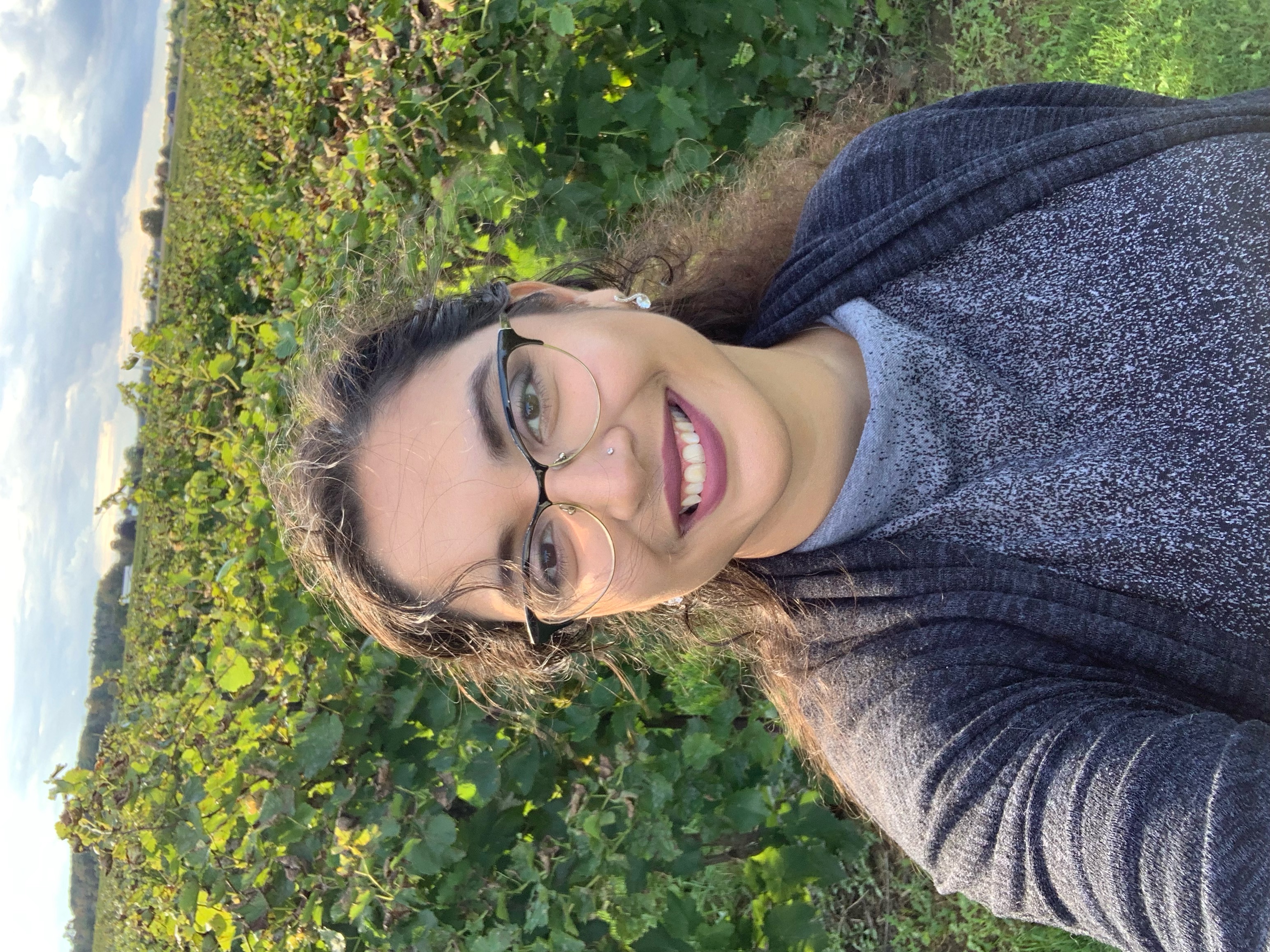
Elizabeth Jewlal
Graduate Student
- Craniofacial Biomechanics & Morphology
- Phenotypic Variation & Variability
Trivia master

Bianca Neale
Graduate Student
- Cranial morphology and evolution
- Skeletal systems
- Squamate evolution
Lover of all reptiles
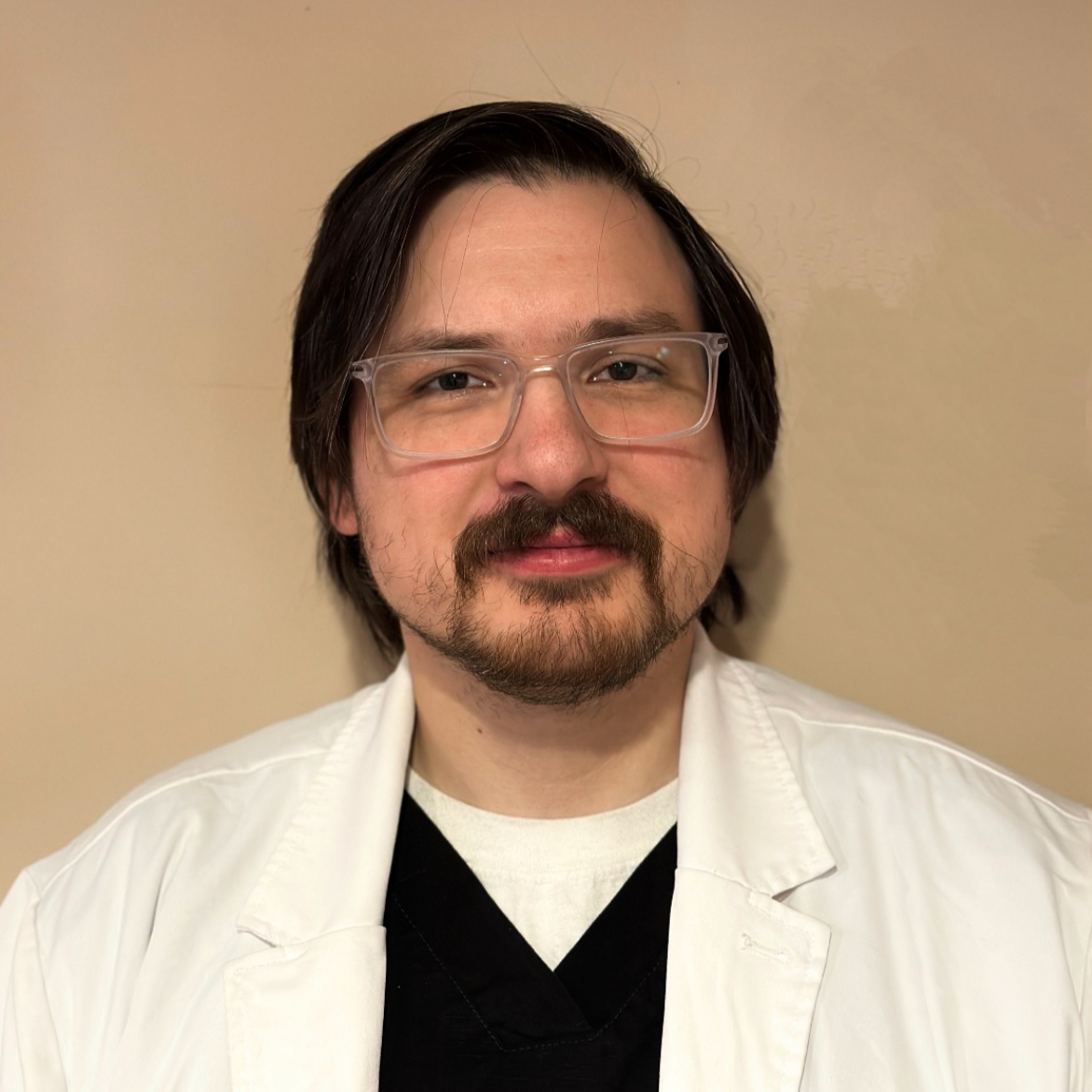
Chris Dulny
Laboratory Technician
Courses Taught
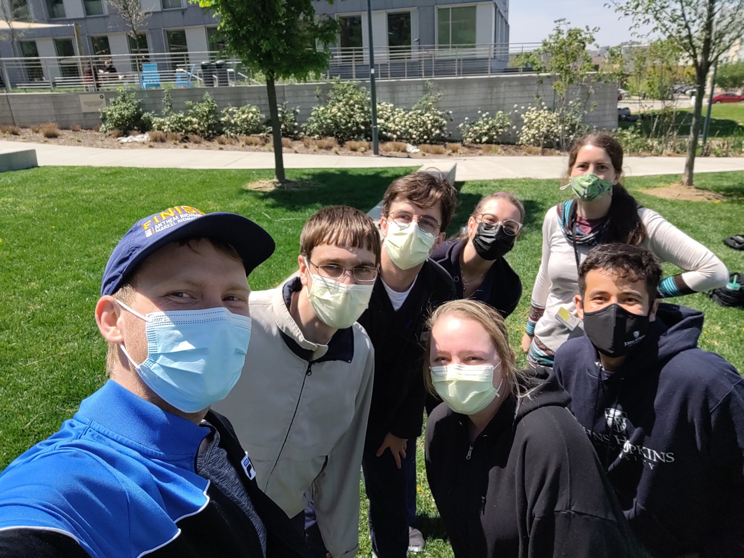
ME.130.753
Fundamentals of Anatomy
This summer course introduces students in the Nurse Anesthetist Doctor of Nursing Practice program to human anatomy using a regional approach. The course is broken into 3 parts – (1) thorax, abdomen, pelvis (2) limbs and back, and (3) head and neck. Within each part, information is presented on the relevant regional topics via: readings and lectures; student observation of prosections in lab; student collaboration to complete model- and computer-based activities.
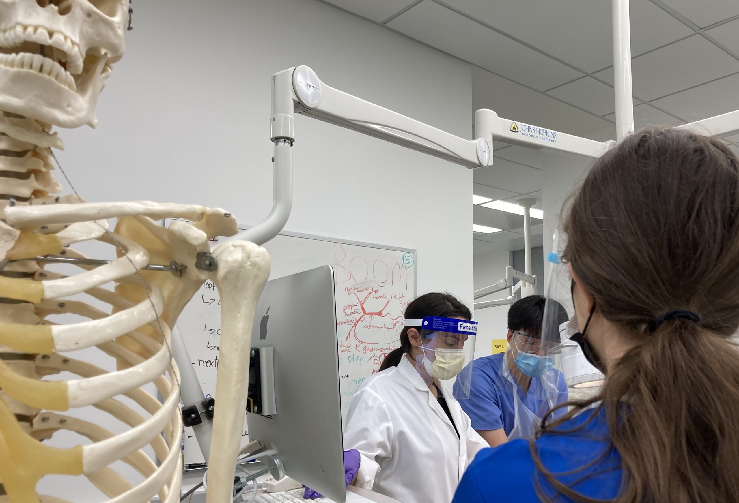
ME: 130.600
SFM Human Gross Anatomy
This seven-week Human Anatomy course is taught to first year medical students in the Johns Hopkins University School of Medicine. Designed to provide a comprehensive regional approach to the human body, this course includes lecture, cadaver dissection with emphasis on the three-dimensional relationships of anatomic structures, clinical correlations, medical imaging sessions, and team-based learning small group activities.
News
FAE Seminar Series presents: Dr. Nicolas Charon
Phd Candidate Zana Sims awarded 2022 Leakey Foundation Grant!
Help us congratulate Zana Sims! Zana Sims was awarded a grant from The Leakey Foundation for her dissertation project, “Examining phylogenetic and dietary signals using cervical root cross sections in extant catarrhines.” Zana's work employs a variety of methods to...

